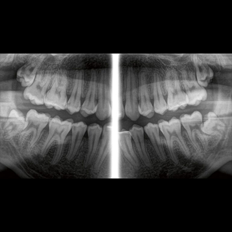
Cephalometric x-rays capture life-sized images of the skull bones and soft tissues from the front, back, and side views. They are commonly used in orthodontics and orthognathic surgery to assist in treatment planning by clearly showing the spatial relationship between the head and facial bones.
Let us call you
Make an Appointment Now
You can get detailed information about your treatment plan by contacting our health consultants.
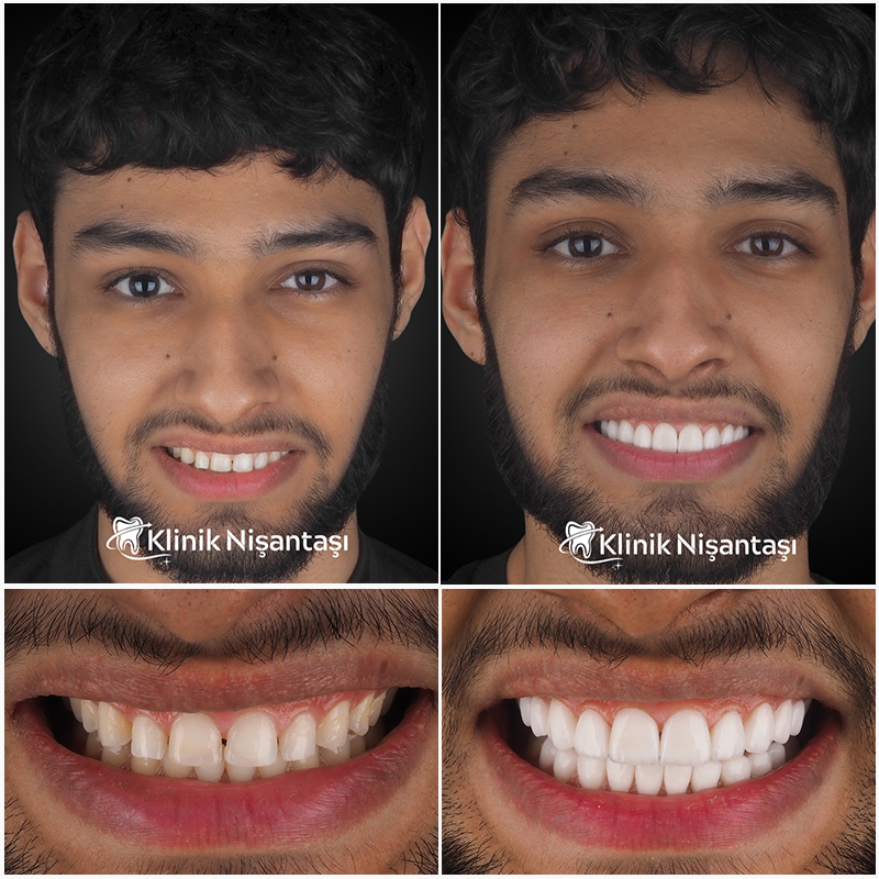
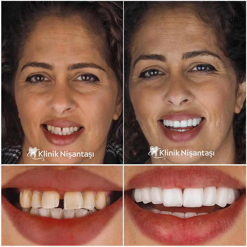
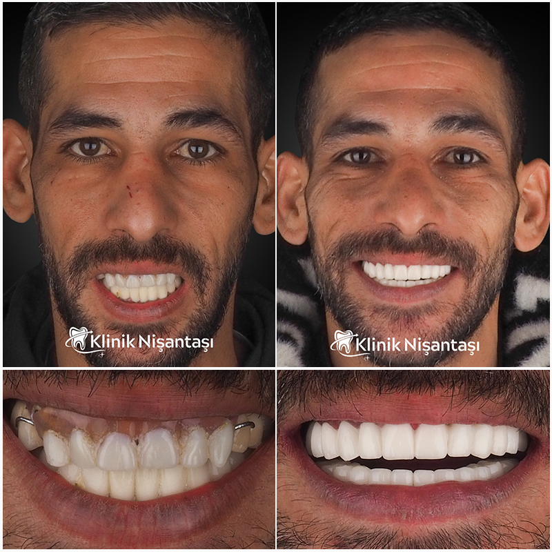
Hear What Our Patients Have to Say About Their Journey
How is a Cephalometric X-ray Taken?
During a cephalometric x-ray, the image sensor is aligned parallel to the patient’s midsagittal plane (the center line dividing the body into left and right). The x-ray source is placed directly opposite the sensor, capturing detailed images of the head bones and soft tissues from the front, back, and side views.
Patient
Experiences
Discover Our Patients Sharing Their Experiences
Bu web sitesi, web deneyiminizi iyileştirmek için çerezleri kullanır.